Comparative Osteology Online
Recommended eBooks
-
 The Bare Bones
by
"An unconventional and reader-friendly introduction to the skeleton as an evolving machine."
The Bare Bones
by
"An unconventional and reader-friendly introduction to the skeleton as an evolving machine."
This 2016 book is the single best title the BPL currently offers access to online for understanding the evolution and functional significance of all parts of the vertebrate skeleton.
Available online through Hoopla. -
 Bone and Muscle
by
A straightforward overview of the functions of the human skeleton and its relationship with musculature.
Bone and Muscle
by
A straightforward overview of the functions of the human skeleton and its relationship with musculature.
Its focus on the human skeleton does not of course obviate its utility for comparative purposes.
Available online through Hoopla. -
 Apes and Human Evolution
by
A 2014 masterwork of comparative primatology that illustrates how effectively skeletal comparisons can be employed to draw conclusions about evolutionary relationships and functional transformations.
Apes and Human Evolution
by
A 2014 masterwork of comparative primatology that illustrates how effectively skeletal comparisons can be employed to draw conclusions about evolutionary relationships and functional transformations.
Available online through Credo Reference.


Related Guides
Introduction
 The comparison of vertebrate skeletons is one of the best ways to understand the ways of life, evolutionary histories, and deep relationships within this group of animals - a group in which we of course have membership.
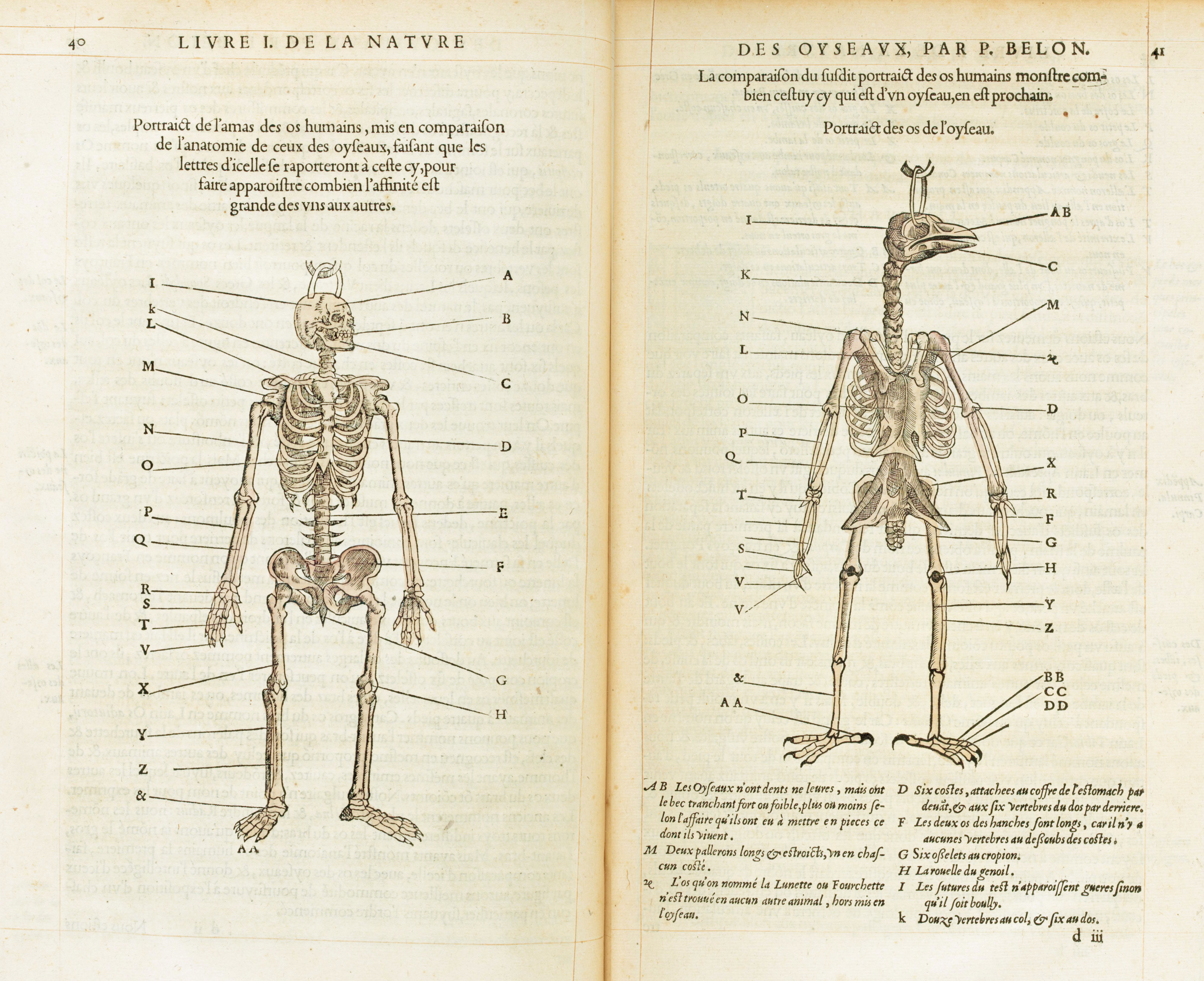
As early as 1555, when Pierre Belon included the illustration at right in his L'Histoire de la nature des oyseaux (Natural History of Birds), it has been recognized that only after stripping away the feathers, scales, fur, and flesh does it become apparent how much apes and birds have in common, and how remarkably similar we are to creatures as different as bats and whales.
This guide organizes an array of resources - all accessible online - to support and encourage the investigation of this fascinating subdiscipline of zoology and anatomy.
Classic Works and Illustrations
Before photography and other imaging technology, the only way to illustrate and communicate evidence from osteological specimens was by drawing. These slides show some examples of incredible skeletal illustration and include links to the original works, all available in digitized form online.
The walrus below comes from Henri-Marie Ducrotay de Blainville's impressively titled Ostéographie, ou, Description iconographique comparée du squelette et du système dentaire des Mammifères récents et fossiles, published over the course of 25 years starting in 1839.
Portions of the complex multipart publication digitized by the Internet Archive are brought together reasonably well by the Biodiversity Heritage Library, and a selection of plates may also be seen at the National Library of Medicine History of Medicine Division's Historical Anatomies on the Web exhibit.

In 1860 Benjamin Waterhouse Hawkins published his Comparative View of the Human and Animal Frame. Hawkins used his artistic skill to create scenes in which skeletons of human and other mammalian species took similar poses, allowing for direct and naturalistic comparison of the bones of the respective species. The image below compares humans and camels. That Hawkins was also a serious anatomist in addition to a great artist is evidenced by the insightful written descriptions that accompany his plates.

Leter, Benjamin Waterhouse Hawkins joined forces with Thomas Henry Huxley, the biologist and famously vigorous supporter of Darwin, to form a sort of osteological superteam, and in 1864 they released the spectacular Elementary Atlas of Comparative Osteology in Twelve Plates. Plate VI, below, depicts an array of vertebrae, beautifully and fearfully sculpted through eons of selection.

Another wonderful plate from Hawkins and Huxley. This one, plate IX, compares the forelimb skeletons of a number of mammals, birds, and non-avian reptiles.

This schematic illustration of a generalized mammal skull from William Henry Flower's 1876 Introduction to the Osteology of the Mammalia resembles something out of a technical manual. Its simplified forms encourage a conceptualization of the mammal skull that allows one to see the affinities and commonalities (derived from common ancestry) underlying the distinct forms of members of the class.

As this illustration of a wolverine skull and teeth shows, Edmond Hue's 1907 Musée ostéologique étude de la faune quaternaire does a particularly good job of representing dentition, portraying its subjects' teeth in an orderly array in both side and top views. The available reproductions were made by Google so the quality is a bit dodgy, but they're still worth a look. Scans may be viewed on HathiTrust.

The left page below from Sidney Reynolds' 1913 The Vertebrate Skeleton makes use of the technique, also used in mechanical drawing, of 'exploding' this green sea turtle skull to render visible portions that would otherwise be obscured in the point of view depicted. This method has been employed using newer visualization technology as well (see below, in 3d Models). A section of this same species' skull appears in the page at right below.

3D Models
The advent of computing brought about the next great wave of osteological visualization in the form of 3d modeling. In some cases, these models are arguably able to convey even more information than would an examination of the physical originals, especially if viewed only in a museum case. This gallery shares some interesting informative innovations used in 3d modeling and provides links to free collections of 3d osteological models online.
Digital Morphology, or DigiMorph, is hosted at UT Austin and is part of the National Science Foundation Digital Libraries Initiative. It is probably the premier online site for 3d visualizations of vertebrate skeletons and skeletal components.
The skull below appears birdlike - but is it a bird? Click on the animation to learn which creature has this curious cranium.
Credit: Dr. Ted Macrini, 2004, "Zaglossus bartoni" (On-line), Digital Morphology. Accessed May 4, 2020 at http://digimorph.org/specimens/Zaglossus_bartoni/.

Other collections may not match DigiMorph in the extent of their holdings or the depth of their accompanying information, but have added features like manipulability and naturalistic surface coloring.
The Osteologic Atlas from the Museu de Ciències Naturals de Barcelona offers a variety of 3d osteological models, primarily of the skulls and mandibles of mammals and birds. The skull below is of a male hammerhead bat.

A brown-throated three-toed sloth skull, also from the Museu de Ciències Naturals de Barcelona, showing the remarkable zygomatic bones (cheekbones), which flare out and back from the forward part of the skull and do not reconnect. The 3d model offers particularly useful perspectives for examining these and other unique formations.

Some institutions create 3d models not only of skulls or other skeletal units, but of entire skeletons. The Idaho Virtual Museum has some examples of this among its collection of 3d models, which is particularly strong in mammals and birds.
The humpback whale whose skeleton was used to create the model below was 44 years old and pregnant when she was struck and killed by a cruise ship in Glacier Bay, Alaska.

This image, also from a model from Idaho Virtual Museum, illustrates the particular utility of the 3d model for examining skeletons. From within the shell of a leopard tortoise, it looks out along the spine towards the skull, showing how the shoulders are suspended from and encased within the shell. 3d modeling makes available lines of sight that might be impossible even when examining a physical specimen in one's hand.

The University of Dundee in Scotland has a collection of 3d models that is kind of a hodgepodge, with some items with wonderful surface detail like this delightfully strange skull of a dugong. This species is the closest living relative of the great Steller's sea cow.

The Museum für Naturkunde Berlin has a wonderful collection of 3d ungulate skull models, like this Abyssinian mohr, a kind of gazelle.

In addition to an excellent collection of amphibian and non-avian reptile models, the Blackburn Lab at the University of Florida's Florida Museum of Natural History also has a fine selection of forelimb models with each bone color-coded for easy comparison between species. Contrasted below are the forelimbs of a snowy egret (left) and a star-nosed mole (right). The bones are (almost) all the same, conserved from a common ancestor, but alterations in relative size and shape result in very different outcomes and utilities.

The Witmer Lab at Ohio University employs some interesting features in their 3d models, including embedded vasculature and brain models and, most engagingly, the use of animation. Below are stills from an animation showing how moveable bones in the upper skull of the scarlet macaw allow for a greater range of motion for its bill through a phenomenon called 'cranial kinesis.' The effect is most pronounced in the animation itself, so click on the image below and have a watch.

In a recapitulation of the method employed by Reynolds with the green sea turtle skull above, the Witmer Lab animation of which stills are shown below 'explodes' the human skull to show its constituent components, reminding us that our skull is in fact made of a large number of individual bones, and in many other species these bones remain functionally distinct and capable of moving in isolation.
